• Equus-Soma

    Equine Osteology & Anatomy Learning Center

    RESEARCH .... DISCOVERY .... EDUCATION

  • Aiken, SC

    info@equus-soma.com

     
  • Waldoboro, ME

    207-542-6132

  • ECVM - Equine Complex Vertebral Malformation

              ECVM is the acronym recently assigned (May-Davis, 2023) to a developmental anomaly affecting the cervical vertebrae and musculature at the base of the horse's neck, specifically the sixth and seventh cervical vertebrae (C6 & C7), the 1st and 2nd sternal ribs and associated soft tissue structures.  
              The presence of this condition was brought to the attention of the horse world by Australian equine anatomist Sharon May-Davis.  Sharon's peer-reviewed publications, resulting from years of research and hundreds of dissections, established the presence of this condition in a variety of breeds, most notably Thoroughbreds and Thoroughbred derivatives. In the past decade, others have published similar findings that support her observations.  Both American and Italian studies found the ECVM in Warmbloods, Quarter Horses and Arabs.  (See Sidebar for links to all references).

  •          Our introduction to this anomaly dates back to 2016 with the recovery of a partial skeleton of a 3 year-old Thoroughbred mare who had the malformation of C6 and C7.  Since then, we have accumulated skeletons from over 50 horses of different breeds and ages.  At the time of this writing 24 of these skeletons had ECVM and each one is morphologically different from the next. Our collection includes several Thoroughbreds, a Friesian, various Warmbloods, a Draught, a few Quarter Horses and 2 Appaloosas plus a few of undetermined breeding.
             Below you will find a summary of what textbooks describe as normal cervical morphology.  The description of what constitutes ECVM relative to C6 can be found here.

  • Cervical Morphology - Overview

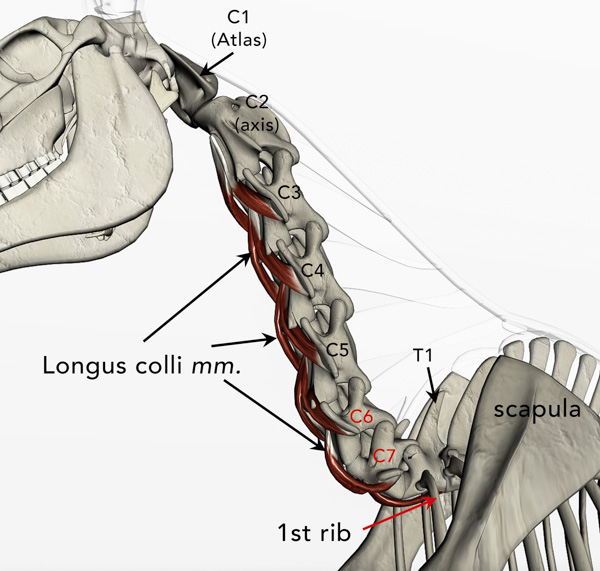
             - Horses, like all mammals (with the exception of sloths and manatees) have 7 neck or "cervical" vertebrae.
             - The first cervical, C1 or "atlas", connects to the base of the skull.
             - Both C1 and C2 ("axis") have distinctly different morphologies from each other, as well as from the remaining cervical vertebrae. 
             - C3, C4 and C5 are generally similar to one-another in appearance but each gets progressively shorter and broader.
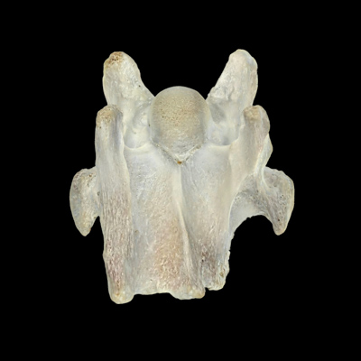
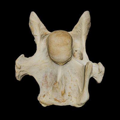
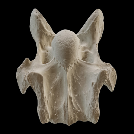
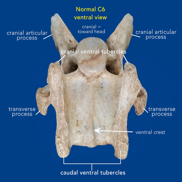
             The ventral side of C6 however, is distinguished from the other cervical vertebrae by the presence of bony "ridges" that extend from the cranial to caudal ends along the left and right sides of the vertebral body (click on image at left).  These "ventral tubercles" or "laminae" are actually branches off of the transverse processes and in the normal C6 are of equal, or near equal, length (cranio-caudally).

  • 3D model of normal C6. Click/tap and drag. Click/tap a number for description.
  •           The last cervical vertebra (C7) is the shortest of all with its ventral surface also differing from that of C6.  The normal C7 does not have the left and  right ventral tubercles characteristic of C6.  Instead, C7 has a pair of small tubercles that lie along the midline instead of a ventral crest (as illustrated by #3 in the 3D model).
              The ventral tubercles of C6 are attachment sights for the strong tendons of the longus colli muscles that control flexion, rotation and stability of the base of the neck (May-Davis, 2015).  These paired, deep perivertebral muscles are highly innervated with proprioceptive characteristics and are considered to be "cybernetic" muscles (Denoix & Pailloux, 2011). The role of cybernetic muscles is to sense motion and orchestrate fine postural movements.   A detailed description of the l. colli musculature can be viewed in this PDF.

  • Sharon May-Davis: Explains ECVM
  • Pamela & Diane Review ECVM Updates